
This story is featured in the Asia Research News 2024 magazine. If you would like to receive regular research news, join our growing community.
Get the news in your inbox
Researchers of RIKEN at Japan’s state-of-the-art synchrotron radiation facility, SPring-8, and their collaborators have developed a faster and simpler way to carry out segmentation analysis, a vital process in materials science. The new method was published in the journal Science and Technology of Advanced Materials: Methods.
Segmentation analysis is used to understand the fine-scale composition of a material. It identifies distinct regions (or “segments”) with specific compositions, structural characteristics, or properties. This helps evaluate the suitability of a material for specific functions, as we ll as its possible limitations. It can also be used for quality control in material fabrication and for identifying points of weakness when analysing materials that have failed.
Segmentation analysis is very important for synchrotron radiation X-ray computed tomography (SR-CT), which is similar to conventional medical CT scanning but uses intense focused X-rays produced by electrons circulating in a storage ring at nearly the speed of light. The team has demonstrated that machine learning is capable of conducting the segmentation analysis for the refraction contrast CT, which is especially useful for visualising the three-dimensional structure in samples with small density differences between regions of interest, such as epoxy resins.
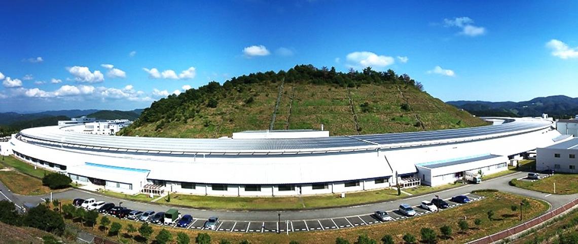
SPring-8 is one of five large synchrotron radiation facilities in the world. SPring-8 stands for Super Photon Ring 8 Gigaelectron volt (GeV), referring to its beam energy of 8 billion electron volts.
“Until now, no general segmentation analysis method for synchrotron radiation refraction contrast CT has been reported,” says first author Satoru Hamamoto. “Researchers have generally had to do segmentation analysis by trial and error, which has made it difficult for those who are not experts.”
The team’s solution was to use machine learning methods established in biomedical fields in combination with a transfer learning technique to finely adjust to the segmentation analysis of SR-CTs. Building on the existing machine learning model greatly reduced the amount of training data needed to get results.
“We’ve demonstrated that fast and accurate segmentation analysis is possible using machine learning methods, at a reasonable computational cost, and in a way that should allow non-experts to achieve levels of accuracy similar to experts,” says Takaki Hatsui, who led the research group.
The researchers carried out a proof-of-concept analysis in which they successfully detected regions created by water within an epoxy resin. Their success suggests that the technique will be useful for analysing a wide range of materials.
To make this analysis method available as widely and quickly as possible, the team plans to establish segmentation analysis as a service offered to external researchers by the SPring-8 data centre, which has recently started its operation.
Further information
Public Relations Office
[email protected]
RIKEN
Dr Yasufumi Nakamichi
[email protected]
Science and Technology of Advanced Materials: Methods (STAM-M)
We welcome you to reproduce articles in Asia Research News 2024 provided appropriate credit is given to Asia Research News and the research institutions featured.